Figure 1.
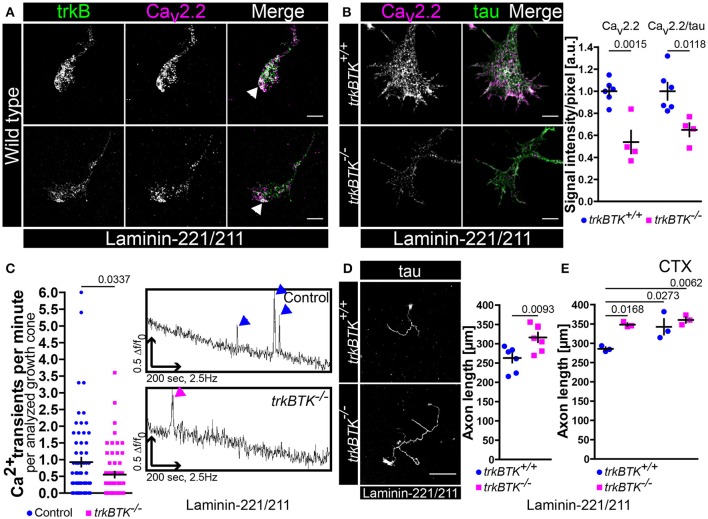
Decrease of Cav2.2 accumulations and spontaneous Ca2+ transients in growth cones of trkBTK−/− motoneurons corresponds to altered axon growth on laminin-221. (A) Representative images of axonal growth cones of wild type motoneurons cultured on laminin-221/211 for 5 days in vitro in the presence of BDNF and CNTF. TrkB receptors (green) and Cav2.2 calcium channels (magenta) were closely localized in growth cone protrusions, highlighted by white arrowheads (scale bar: 5 μm). (B) In trkBTK−/− growth cones Cav2.2 accumulation (magenta) was affected in growth cone tips, whereas tau levels (green) were not altered (scale bar: 5 μm). Statistical analysis of Cav2.2 immunoreactivity in trkBTK−/− axonal growth cones (0.54 ± 0.1, Q2 0.47, n = 4, N = 66) in comparison to wild type controls (1.00 ± 0.04, Q2 1.01, n = 6, N = 78) revealed a significant difference (p = 0.0015). Similar findings were obtained by normalizing Cav2.2 against the internal reference protein tau (trkBTK+/+ 1.00 ± 0.08, Q2 0.95; trkBTK−/− 0.65 ± 0.06, Q2 0.67; p = 0.0118) (C) The structural phenotype was accompanied by significant differences in the frequency of spontaneous calcium transients between control and trkBTK−/− growth cones (Control 0.92 ± 0.13, Q2 0.60, N = 72; trkBTK−/− 0.55 ± 0.09, Q2 0.30, N = 64; p = 0.0337). Representative recordings of control (upper trace, blue arrowheads) and trkBTK−/− (lower trace, magenta arrowheads) growth cones showed a reduced number of calcium spikes when trkB signaling is impaired. (D) Wild type and trkBTK−/− motoneurons were cultured on laminin-221/211 for 7 days in vitro in the presence of BDNF and CNTF and stained against tau (scale bar: 150 μm). TrkB mutant cells (316.4 ± 12.65 μm, Q2 314.5 μm, n = 7, N = 298) grew longer axons than controls (262.9 ± 11.79 μm, Q2 278.8 μm, n = 7, N = 283, p = 0.0093) (E) Treatment with 30 nM ω-conotoxin (CTX) caused enhanced axonal elongation in wild type motoneurons, but not in trkBTK−/− cells.

