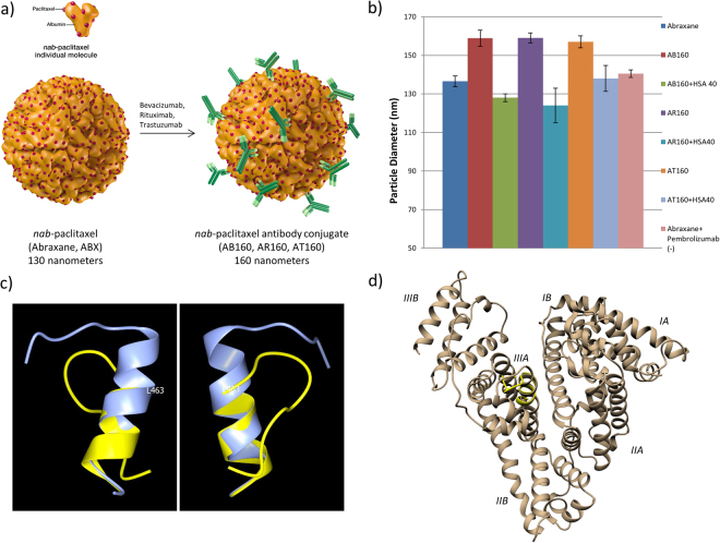
Figure 2.
Monoclonal Antibody Coated Nab-Paclitaxel Nanoparticle Formation Inhibition by HSA Peptide 40. (a) Particle formation schematic, 130 nm nab-paclitaxel (Abraxane) nanoparticles are incubated with either bevacizumab (Avastin), rituximab (Rituxan), trastuzumab (Herceptin), or pembrolizumab (Keytruda) to form 160 nm antibody directed nanoparticles AB160, AR160, AT160, and a negative control Abraxane + Pembrolizumab. (b) 10 mg/ml of Abraxane was incubated for 30 minutes with 4 mg/ml of either bevacizumab, rituximab, or trastuzumab to test formation of AB160, AR160, or AT160 in the presence of a molar excess of HSA Peptide 40. Particle sizes were determined by NS300 dynamic light scattering and nanoparticle tracking system. Blocking of formation of 160 nm particles suggest HSA Peptide 40 outcompetes the in-particle albumin for the binding site on the antibodies. 3 repeats were performed for each experiment and error bars represent the standard error of measurement determined by Nanosight NTA 3.1 Software. (c) Superposition of predicted HSA peptide 40 model (yellow) onto Val-455 – Arg-472 in HAS X-ray crystal structure 1AO6 (blue). Structures drawn in ribbon representation. Two views approximately 180° apart. Leu-463 in 1AO6 labeled. (d) HSA Pep 40 (yellow) superimposed onto human serum albumin using MatchMaker, HSA domains and subdomains labeled.

