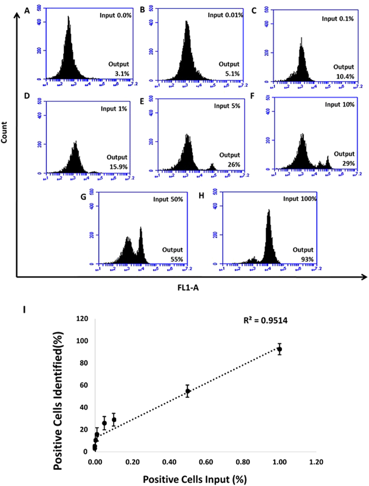
Figure 11.
Selective recognition of MCF7 cells in mixed cell samples with the 6-FAM labelled MAMB1 aptamer in whole blood lysate. Cell mixture samples containing MCF7 and PBMC were prepared in different percentages of MCF7 ((A–G) 0%, 0.01%, 0.1%, 1%, 5%, 10%, 50%, and 100%). (I) plot of the percentage of cancer cells (MCF7) spiked (X-axis) and the percentage of positive cells identified (Y-axis). Values are shown as means ± S.E.M. of three trials. The statistical significance was determined by one way ANOVA followed by Fisher LSD multiple tests using SPSS software (SPSS, version 23). P Values less than 0.05 were considered to be significantly different.

