Figure 2.
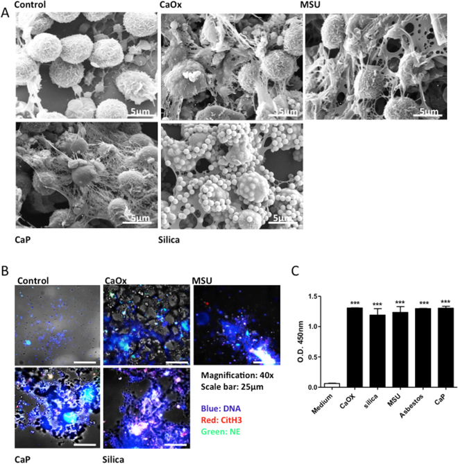
Different crystals induce NET formation. Human neutrophils were exposed to crystals of calcium oxalate (CaOx) (0.2 mg/ml), MSU (0.2 mg/ml), calcium phosphate (0.2 mg/ml), and silica (0.2 mg/ml) for 2 hours. Scanning electron microscopy was used to visualize crystal associated NETs (scale bar = 5 µm). Please note some platelets alongside the neutrophils in the control image. Arrows indicate the presence of NETs and NET-crystal aggregates. (A) Neutrophils exposed to crystals were co-stained for NET markers DNA (blue), citrullinated histones H3 (red) and neutrophil elastase (green). Cells were imaged using a fluorescence microscope. Crystals can be seen in phase contrast (grey) (magnification 40x). (B) NETs were quantified by MPO-DNA complex ELISA after 2 hours of crystal exposure (C). All representative images are from a single experiment. Data were obtained from three independent experiments each performed in duplicate. Data represent means ± SEM. ***p < 0.001 versus medium.

