Figure 3.
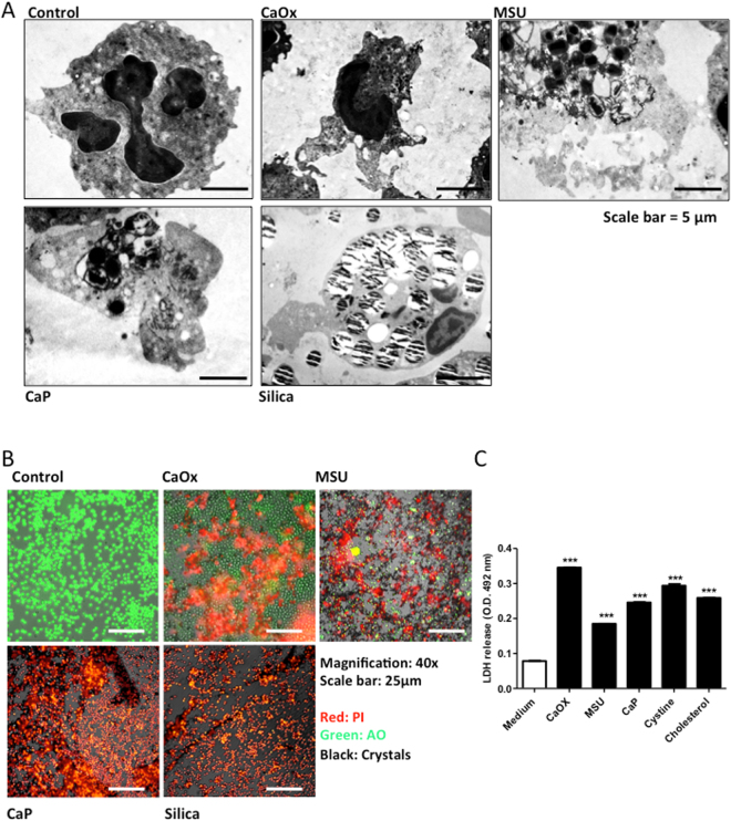
Different crystals induce neutrophil cell death. Human neutrophils were exposed to crystals of calcium oxalate (0.2 mg/ml), MSU (0.2 mg/ml), calcium phosphate (0.2 mg/ml), and silica (0.2 mg/ml) for 2 hours. Transmission electron microscopy was used to visualize crystal-associated neutrophil cell-death (scale bar = 5 µm) (A). Crystal-induced cell death was visualized by live cell imaging using propidium iodide (PI, red area). Acridine orange (AO) stained live cells. Crystals were imaged in phase contrast (grey) (magnification 20x) (B). LDH release was measured in the supernatants of crystal-exposed neutrophils post 2 hours (C). All representative images are from a single experiment. Data were obtained from three independent experiments each performed in duplicate. Data represent means ± SEM. **p < 0.01, ***p < 0.001 versus medium.

