Figure 2.
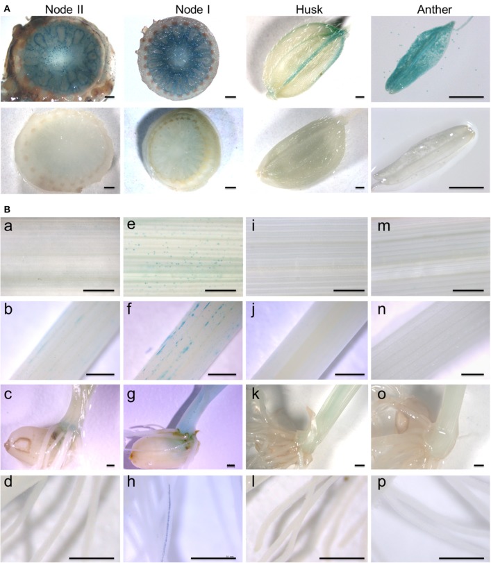
Spatial and temporal expression of the OsARM1pro::GUS construct. (A) Histochemical GUS staining of transgenic rice expressing OsARM1pro::GUS showing high levels of GUS signal in the enlarged vascular bundles of node I and node II, the vessel tissues of the husk, and the anther (upper images). GUS staining of wild-type SSBM was used as a negative control (bottom images). (B) The expression patterns of OsARM1pro::GUS upon exposure to As(III). Various organs were collected from two-week-old seedlings at the vegetative growth stage. The OsARM1pro::GUS seedlings were treated with 50 μM As(III), and the samples were harvested at 0 and 6 h after treatment. Images in (e–h) show As(III)-treated samples of leaves (e), stems (f), basal regions (g), and roots (h) of OsARM1pro::GUS seedlings. Images in (a–d) show the corresponding untreated controls. Wild-type SSBM samples harvested from As(III)-treated (m–p) or untreated (i–l) plants were used as negative controls. Bars = 500 μm.

