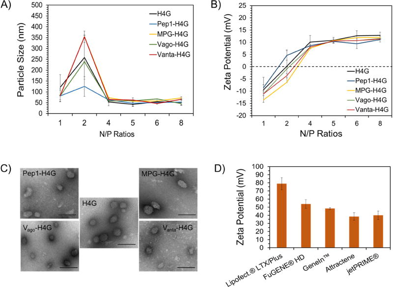
Figure 3.
Characterization of nanoparticles in terms of size, charge, and shape. A) Size of DBV/pEGFP nanocomplexes as determined by dynamic light scattering. B) Surface charge of DBV/pEGFP nanocomplexes as determined by laser Doppler velocimetry. C) Shape of DBV/pEGFP nanocomplexes captured by TEM. The scale bar is 100nm (magnification: 75000×). D) Surface charge analysis of commercial vectors in complex with pEGFP.

