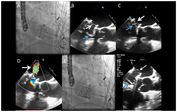
Fig. 1.
Transcatheter mitral valve-in ring. A: A 26-mm Edwards Sapien valve was deployed at the level of the mitral valve ring using a transapical approach. B: Intraoperative transesophageal echocardiogram (TEE) after mitral valve-in-ring replacement revealed native valve leaflets (blue arrow) overhanging and interfering (C) with closure of the prosthetic leaflets (white arrow), and resulting severe central MR (D) by color Doppler (arrow). D: A 22-mm Melody valve was deployed farther into the atrium away from the prolapsing leaflet. E: TEE after second transcatheter valve showing functioning transcatheter valve leaflets (white arrow) that are a significant distance from native valve leaflets (white arrow). [Color figure can be viewed at wileyonlinelibrary.com]

