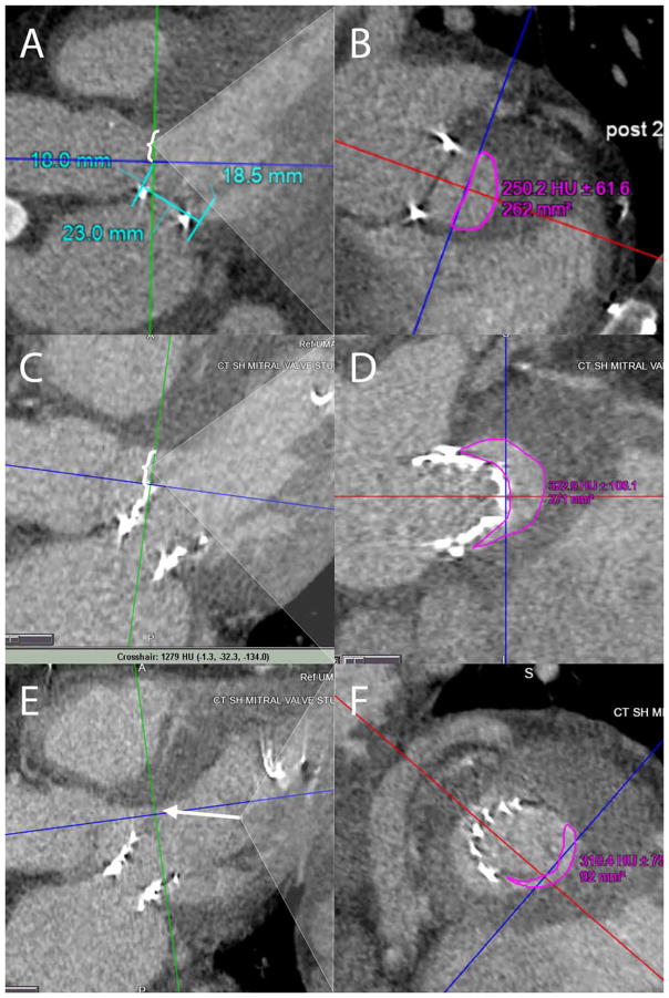
Fig. 3.
A, B: Pre-procedural computed tomography (CT) analysis was used to simulate the 23-mm Sapien 3 valve superimposed in the mitral space and the cross-sectional area remaining in the left ventricular outflow tract (LVOT) area was estimated (bracket) at 262 mm2 in the green plane depicted in panel (B). C, D: Post-Sapien 3 implantation CT LVOT measurement (bracket) demonstrated an adequate area of 271 mm2 when measured at the Sapien 3 stent frame. However, since the “neo-LVOT” comprises of the stent frame and anterior mitral leaflet, it was more accurate to measure the “neo- LVOT” at the tip of the anterior mitral leaflet (arrow). This “neo-LVOT” measured 92 mm2 (E and F), which could explain the flow acceleration in the LVOT after preload reduction from a dialysis session. [Color figure can be viewed at wileyonlinelibrary.com]

