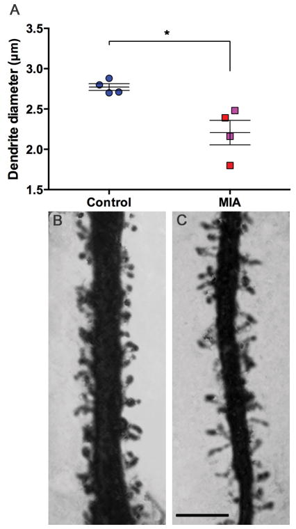
Figure 4.
Diameter (μm) of apical dendrite shaft. MIA animals have thinner apical dendrites than control animals (A). Representative photomicrographs of control (B) and MIA (C) apical dendrites. (Control animals (all male – (blue)), MIA males (red), MIA female animals (pink)), scale bar = 5μm, *P<0.05)

