
FIGURE 3.
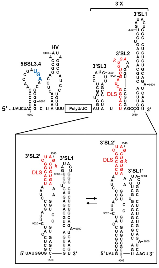
The 3′UTR of the HCV RNA genome. Secondary structure model proposed for the 3′UTR, showing the theoretical alternative conformations acquired in the 3′X tail. The palindromic motif involved in viral genome dimerization (DLS, dimer linkage sequence) is shown in red. The k sequence required for the interaction with the apical loop of the 5BSL3.2 domain is shown in italics. The translation stop codon is marked in enlarged blue text. Nucleotide numbering is as in Figure 2.