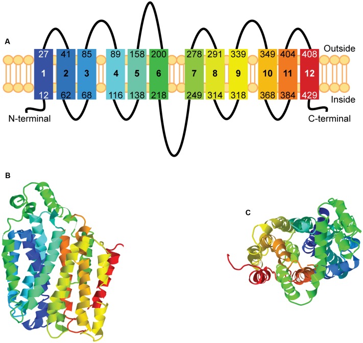
FIGURE 2.
Homology modeling of hsUNC93A. The secondary and tertiary structures of hsUNC93A were modeled using Phyre2. The secondary structure (A) of UNC93A revealed 12 transmembrane helices, marked by 1–12, similar to other MFS proteins. The prediction of the tertiary structure illustrated a globular protein (B, side-view), with helices packed to form a pore (C, top-view). The rainbow gradient of the secondary and tertiary structures shows helices from N-terminal (dark blue) to C-terminal (red).

