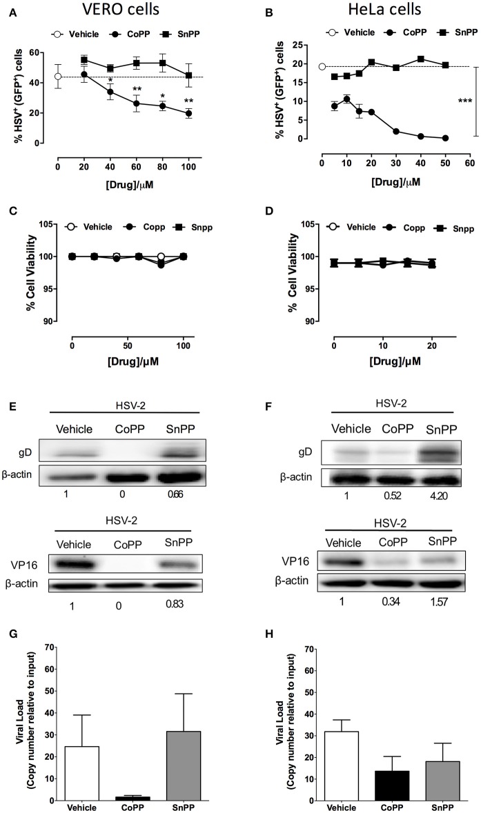
Figure 2.
Pharmacological induction of HO-1 restricts the expression of HSV-encoded genes in epithelial cells. (A,B) Flow cytometry analyses of GFP-derived fluorescence in Vero (left) and HeLa cells (right), respectively infected at MOI 1 with HSV-2 that encodes a non-structural GFP reporter gene under the control of a constitutive promotor, in response to varying concentrations of HO-1-modulating drugs. (C,D) Cell viability was assessed for Vero (left) and HeLa cells (right) for treatment with varying concentrations of HO-1-modulating drugs. (E,F) Western blot analyses for viral proteins gD and VP16 in Vero (left) and HeLa (right) cells at 16 h post-infection with HSV-2 at an MOI 1 after treatment with 60 and 10 μM, respectively of HO-1-modulating drugs. (G,H) Quantification of viral genome copies in Vero (left) and HeLa cells (right) by qPCR at 18 h post-infection at MOI 10. Data are means ± SEM of three independent experiments for experiments (A–F) and two independent experiments for (G,H). Representative images are shown for Western blots. Two-way ANOVA, and Tukey's multiple comparison test were used for statistical analyses (A): statistics is shown for the CoPP treated group vs. SnPP-treated and untreated (*p < 0.05, **p < 0.01, ***p < 0.001).

