FIGURE 5.

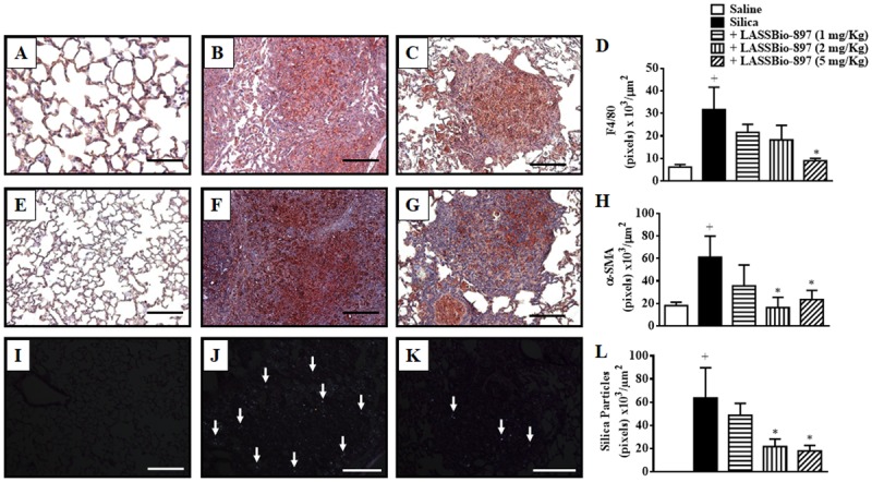
LASSBio-897 reduces macrophages, myofibroblasts, and silica particles into the lungs of silicotic mice. Silica (10 mg) was given i.n. and analyses were performed 28 days later. Oral treatment with LASSBio-897 was performed daily for seven consecutive days, starting 21 days after induction of silicosis. Panels show representative photomicrographs of lung sections of F4/80 immunoreactivity (upper panels), α-SMA immunoreactivity (middle panels), and silica particles (lower panels) of saline-provoked mice (A,E,I); silica-provoked mice (B,F,J); silica-provoked mice treated with LASSBio-897 (5 mg/kg) (C,G,K). (D) and (H) represent the quantification of pixels associated with F4/80 and α-SMA expression, respectively. (L) Quantitative analysis of silica particles. Silicotic non-treated animals received an equal amount of vehicle (DMSO 0.1%). White arrows indicate silica particles in (J) and (K). Data are expressed as the means ± SD from six mice. Scale bars = 200 μm. These results are representative of two independent assays. +P < 0.05 compared to saline-provoked group. ∗P < 0.05 compared to silica-provoked group.
