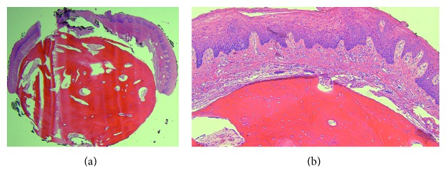
Figure 2.
Lingual osseous choristoma. (a) Low-power photomicrograph demonstrating a nodule of dense cortical lamellar bone underlying benign-appearing stratified squamous epithelium, H&E ×20. (b) Despite its improper location within the subepithelial tissue of the tongue, the bone appears histologically unremarkable with a normal distribution of osteocytic lacunae and haversian canals, H&E ×100.

