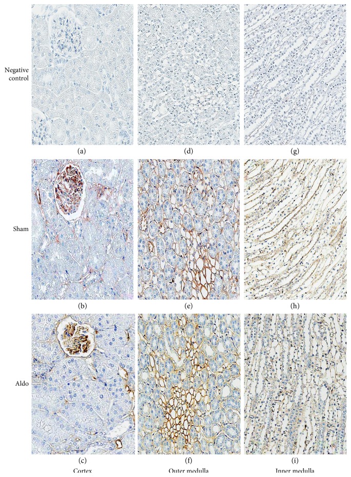
Figure 4.
Representative immunohistochemical staining micrographs of renal PKCβII protein localization in the cortex (a–c), the outer medulla (d–f), and the inner medulla (g–i) from sham (b, e, and h) and Aldo (c, f, and i) (n = 5/group). Negative controls (a, d, and g). Original magnification, ×400 (a–c) and ×200 (d–i).

