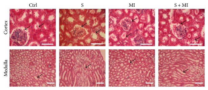
Figure 4.
Effect of S and MI on kidney histopathology, using trichrome staining. The trichrome stain confirmed the absence of glomerulosclerosis and interstitial fibrosis in the smoking (S) group. However, the MI group demonstrated swelling of the PCT cells and rare hyaline casts, identified in the tubular lumen. On the other hand, the S+MI group showed PCT dilation with thinning of the epithelial lining and swelling of the cells. Results are representatives of five different mice in each group (n = 5). Scale bars are 50 μm.

