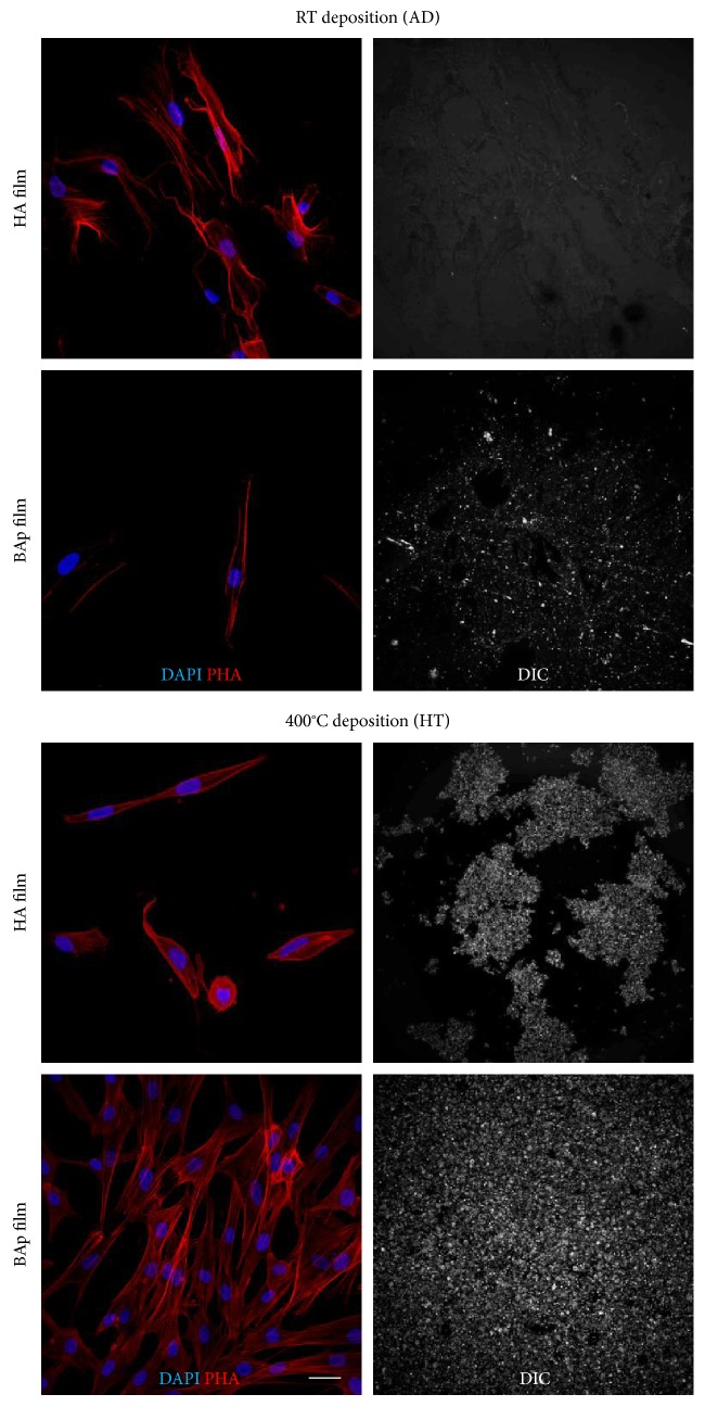
Figure 3.
Representative immunofluorescence images showing cell morphology of hDPSCs cultured on HA and BAp films and DIC images representing film adhesion to the underlying surface at 7 days of culture. Immunofluorescence analysis was performed against phalloidin. Nuclei were counterstained with DAPI. Scale bar: 10 μm.

