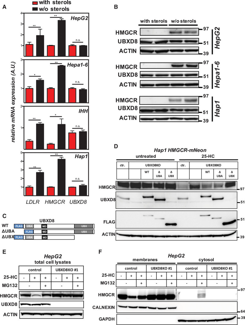
Figure 4.

UBXD8 (ubiquitin regulatory X domain–containing protein 8) promotes degradation of HMGCR (3-hydroxy-3-methylglutaryl coenzyme A reductase) in a UBX domain–dependent manner. A, Hap1, and the hepatocyte lines HepG2, Hepa1-6, and IHH were cultured for 24 h in sterol-containing (red bars) or sterol-depletion medium (black bars). Subsequently, (A) total RNA was isolated, and expression of the indicated genes determined by quantitative polymerase chain reaction (n=3), *P<0.05, **P<0.01, or (B) total cell lysates were immunoblotted as indicated. Immunoblots are representative of 3 independent experiments. C, Schematic representation of FLAG-tagged full length wild type (WT) UBXD8 and its variants lacking the UBA or UBX domain; membrane-associating domain (MD). D, Control Hap1 or Hap1-UBXD8KO cells in which the FLAG-tagged full length or indicated variant were stably reintroduced were cultured in sterol-depletion medium for 24 h and then treated with 10 μmol/L 25-hydroxycholesterol (25-HC) for 1 h as shown. Total cell lysates were immunoblotted as indicated and are representative of 3 independent experiments. E, HepG2 control and UBXD8KO cells were cultured in sterol-containing or sterol-depletion medium for 24 h and subsequently treated with 10 μmol/L 25-HC or 25 μmol/L MG132 for 2 h as shown. Total cell lysates were immunoblotted as indicated and are representative of 3 independent experiments. F, HepG2 control and UBXD8KO cells were treated as described in (E), and membrane and cytosolic fractions were prepared, as described in Materials and Methods in the online-only Data Supplement. Subsequently, 20 or 60 μg protein per lane of the membrane and cytosolic fractions were analyzed by immunoblotting as indicated. Immunoblot is representative of 3 independent experiments. Note that the membrane and cytosolic HMGCR immunoblot are from different exposures.
