Figure 4.

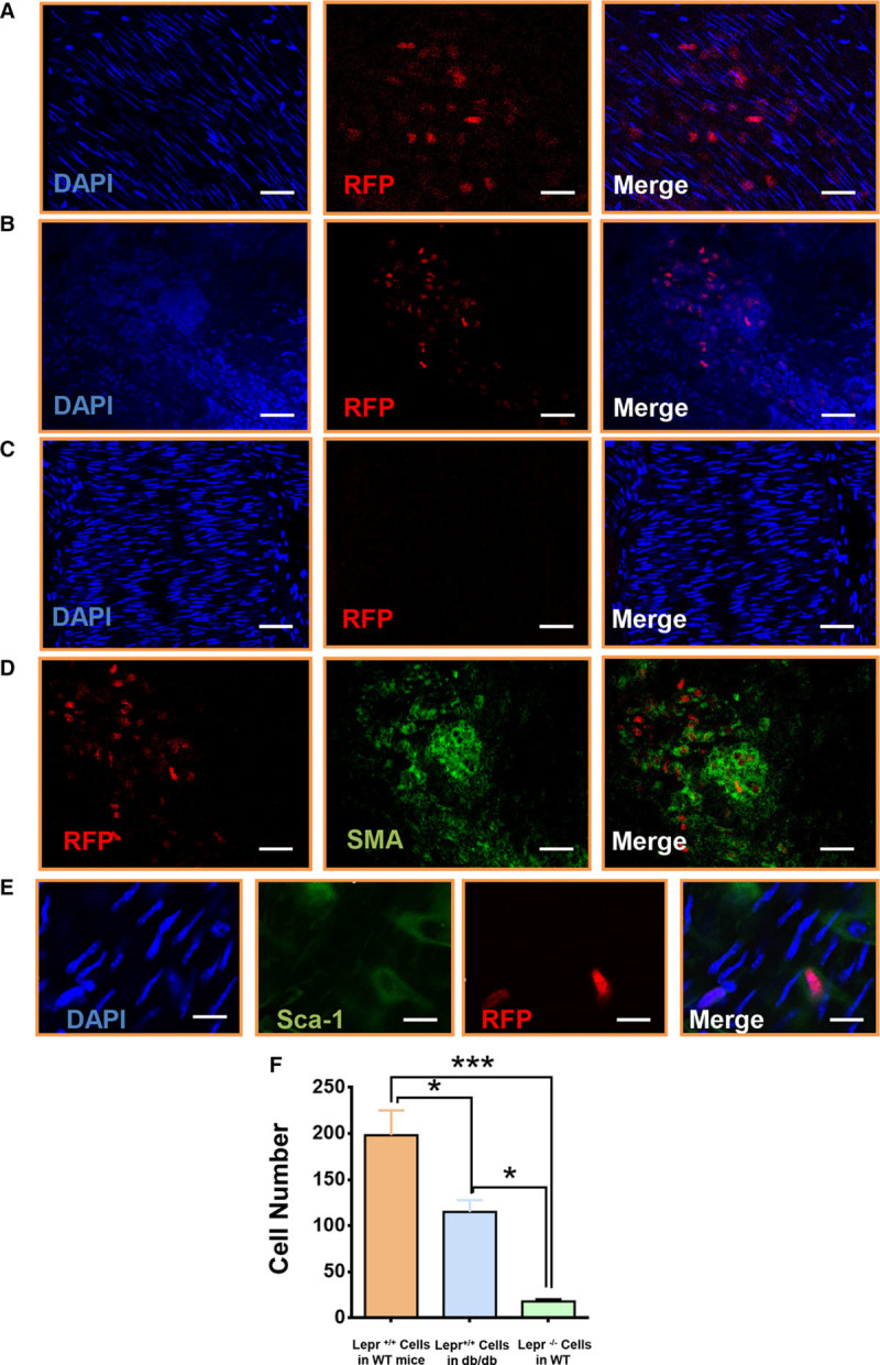
Lack of leptin receptor in Sca-1+ progenitor cells abolished the leptin-induced cell migration in vivo. A, Cell migration assay in vivo was performed in wild-type mice. 1×10⁶ RFP (red fluorescent protein) Sca-1+ were seeded in the adventitial side of injured femoral artery in wild-type mice. En face staining showed that the RFP cells migrated toward the intima at 72 h after the surgery (scale bar, 30 μm; n=6). B, 1×10⁶ RFP Sca-1+ were seeded in the adventitial side of injured femoral artery in leptin receptor–deficient (db/db) mice. En face staining showed that the RFP cells migrated toward the intima at 72 h after the surgery (scale bar, 100 μm; n=6). C, 1×10⁶ RFP lepr−/− Sca-1+ were seeded in the adventitial side of injured femoral artery in wild-type mice. En face staining showed that the RFP cells did not migrate toward the intima at 72 h after the surgery (scale bar, 50 μm; n=6). D, 1×10⁶ RFP Sca-1+ cells were seeded in the adventitial side of injured femoral artery in db/db mice. En face staining showed that the RFP cells acquired smooth muscle cell marker (Alexa 488; green) at 72 h after the surgery (scale bar, 20 μm; n=6). E, 1×10⁶ RFP Sca-1+ cells were seeded in the adventitial side of injured femoral artery in wild-type mice. En face staining showed that some RFP cells lost Sca-1+ marker (Alexa 488; green) at 72 h after the surgery (scale bar, 5 μm; n=6). F, Quantification of migratory cells from the adventitia for group A, B and C. All graphs are shown as mean±SEM. *P<0.05, **P<0.01, ***P<0.001.
