Figure 5.

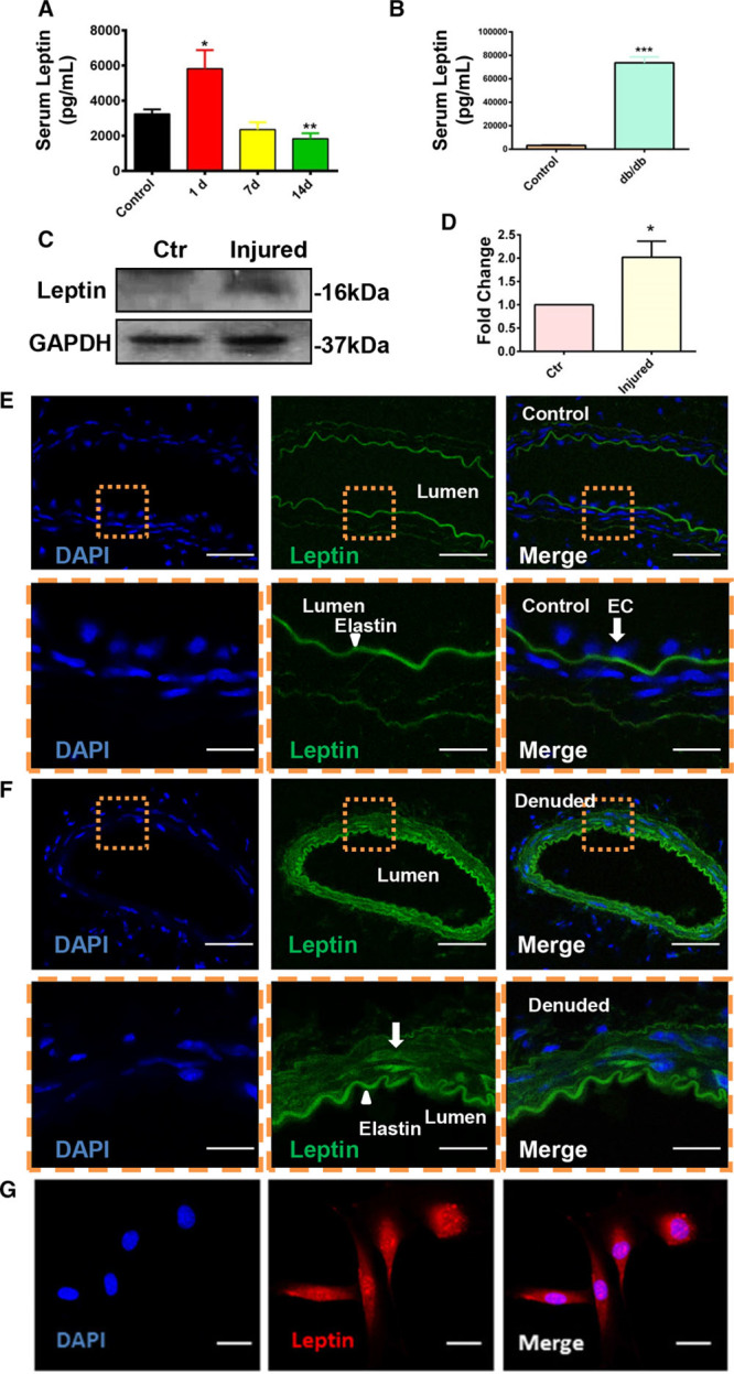
The expression of leptin was upregulated after surgery both in the circulation and in the injured artery. A, Serum leptin in wild-type mice 1 (n=10), 7 (n=5) and 14 (n=12) d after injury were quantified by using leptin ELISA kit. B, Serum leptin in wild-type and db/db mice was quantified by using leptin ELISA kit. C, Expression of leptin in the injured or noninjured arteries was documented by performing Western blotting 1 d post-surgery (n=4). D, Quantification of leptin expression in noninjured and injured vessels. E and F, Difference in expression of leptin (Alexa 488; green) in noninjured (E) artery (n=6) and injured (F) artery (n=10) was analyzed on 1 d post-surgery by immunofluorescence (scale bars, 30 μm). G, Expression of leptin in smooth muscle cell (Alexa 594; red) in vitro was detected by immunofluorescence (scale bars, 8 μm). DAPI indicates 4’,6-diamidino-2-phenylindole for nucleus staining; and EC, endothelium. All graphs are shown as mean±SEM. *P<0.05, **P<0.01, ***P<0.001.
