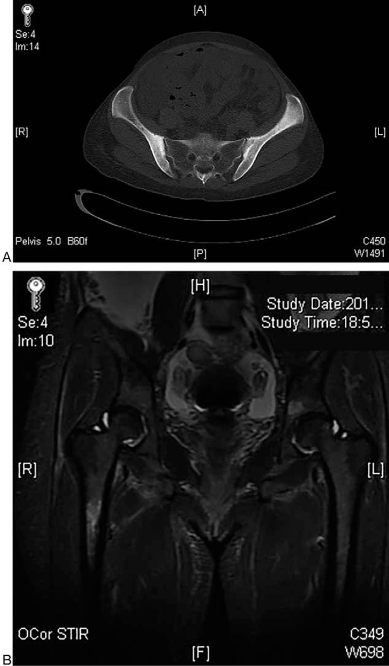
Figure 1.

Imaging studies. (A) The ilia show mildly expansile change with thickened and blurred cortex on contrast-enhanced computed tomography (CT) scan, which indicates ossification and bilateral sacroiliitis, greater on the left. (B) Edema of bone marrow was observed in the right proximal femur, which manifested as increased signal intensity in the short tau inversion recovery (STIR) images.
