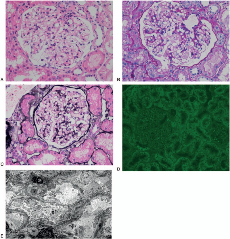
Figure 2.

Renal biopsy. Histological analysis highlighted the thickening and stiffness of the capillary basement membrane (A, Hematoxylin and eosin staining, X400; B, Periodic acid Schiff, X400). PASM staining showed vacuoles in the thick capillary wall without significant spikes (C, X400). PLA2R staining was negative (D, X400). Electron microscopy showed subepithelial granular electron-dense deposits, with some even penetrating the basement membrane. Overlying podocyte foot processes effaced obviously (E).
