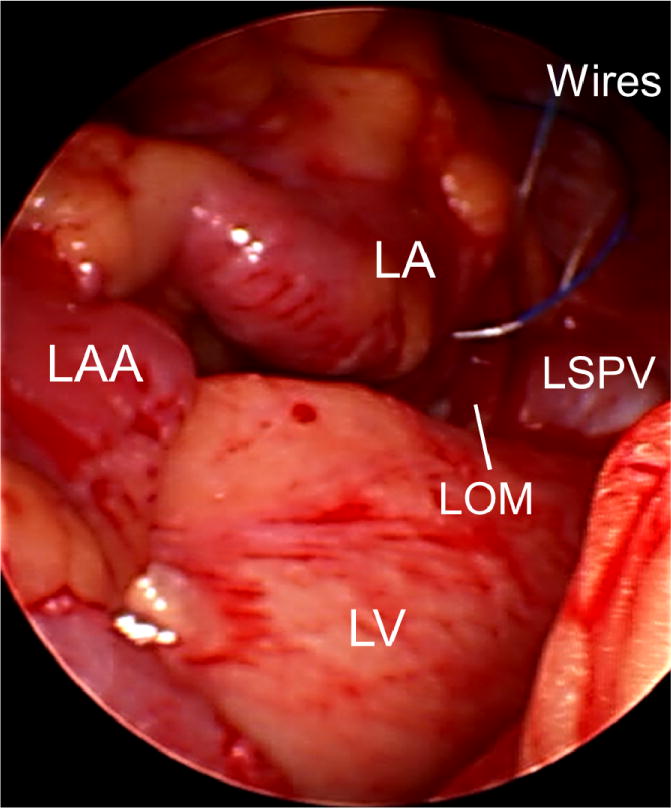
Figure 1.

Placement of the temporary pacing wires during surgery. At the end of the open heart surgery, two temporary pacing wires were threaded through the epicardial fat pad at the junction of the left superior pulmonary vein (LSPV) and left atrium (LA). The uninsulated portion of the temporary pacing wires went through the fat pad (yellow) near the LOM and the orifice of the LSPV. LAA, left atrial appendage; LOM, ligament of Marshall; LV, left ventricle.
