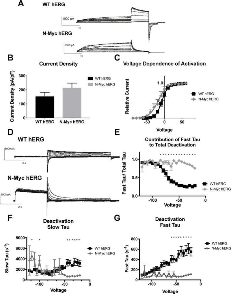
Figure 3. Effects of N-terminal-myc hERG on channel activation and deactivation.

A) Whole cell patch clamp traces of WT hERG and N-terminal-myc hERG activation. Voltage clamp protocol shown below trace. B) Histogram representation of the current density for WT hERG and N-terminal-myc hERG, normalized to cell capacitance. There are no significant differences in current density (n= 12 (WT) and 6 (N-terminal-myc)). C) Representation of Voltage Dependent Activation (VDA) of WT hERG and N-terminal-myc hERG. VDA of N-terminal-myc hERG is hyperpolarized significantly compared to WT hERG. V1/2 of WT hERG is −7.182 ± 0.1371 and the V1/2 of N-terminal-myc hERG is −17.72 ± 0.9231. D) Whole cell patch clamp traces of WT hERG and N-terminal-myc hERG deactivation. Voltage clamp protocol shown below trace. N= 12 (WT hERG) and 8 (N-terminal-myc). E) Contribution of Fast Tau to total time constant of deactivation. There are significant differences from −20 to −85 mV (p<0.05), indicating that N-Myc Fast Tau contributes significantly more to total deactivation. F) Graphical depiction of WT and N-Myc hERG Slow Tau deactivation. There are significant differences at voltages from −20 to −40 and at −100 and −115 (p<0.05), although these may be less relevant differences. G) Representation of Fast Tau deactivation. There are significant differences between WT hERG and N-terminal-myc hERG at voltages from −20 – −60 (p<0.05). All results demonstrate that N-terminal-myc hERG deactivates significantly faster than WT hERG.
