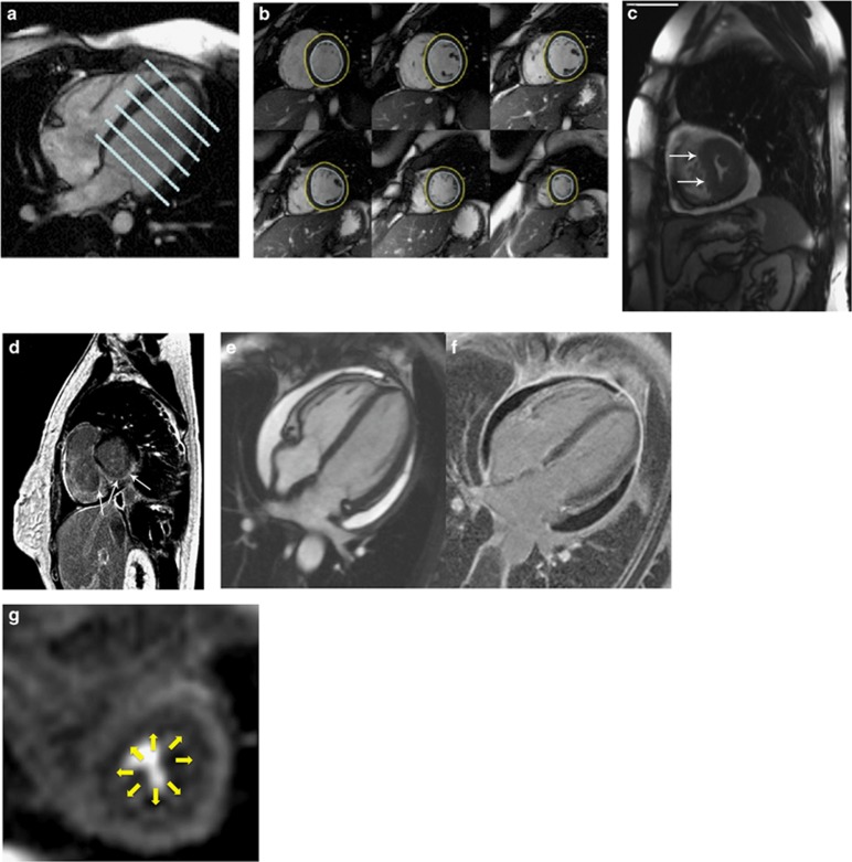
Figure 1.
(a, b) Cine CMR: A four chamber view (left) with positions of each slice of a short axis stack (right). Imaging is performed in the 2-chamber, 3-chamber and 4-chamber long axis views and in a stack of short axis slices, typically acquired every 10 mm from the mitral valve plane to the apex. Mass and volume are quantified from the short axis stack according to Simpson’s Rule by slice summation, which avoids making any geometric assumptions about the shape of the ventricle. The high blood-myocardial contrast and ability to quantify volumes without geometric assumptions is especially useful in the RV, because of its complex shape, abundant trabeculations and thin wall. Because gadolinium contrast agents are confined to the extracellular space, the change in T1 relaxation time is inversely related to the contrast agent volume of distribution. Even without the administration of contrast, native T1 mapping detects prolongation of T1 in SSc. Active inflammation can be assessed by T2-weighted images, which demonstrate increased signal in areas of myocardial edema. Abnormal myocardial T2 signal has been described in patients with SSc.50 Endocardial (blue) and epicardial (yellow) borders are drawn, enabling calculation of left ventricular mass and chamber volume according to Simpson’s rule.55 (c) Diastolic flattening of interventricular septum, ‘D-sign’, visualized by cine CMR. Adapted from Burt et al.22 http://www.jrheum.org/content/39/2/206.long, reproduced with permission from Journal of Rheumatology and Richard Burt. (d) Focal fibrosis in a SSc patient involving the mid and epicardial myocardium, sparing the subendocardium, as visualized by LGE-CMR. Because gadolinium contrast agents are confined to the extracellular space, the change in T1 relaxation time is inversely related to the contrast agent volume of distribution. CMR measurement of the relative gadolinium concentration in the blood and myocardium, and the direct measurement of the volume of distribution of gadolinium in the blood (1-hematocrit), enables calculation of extracellular volume fraction (ECV), an index of the volume of distribution of contrast agent in the myocardium. Adapted from Burt et al.22 http://www.jrheum.org/content/39/2/206.long,20 reproduced with permission from Journal of Rheumatology and Richard Burt. (e, f) Pericardial effusion seen as bright white on Cine CMR (left) and dark black on phase sensitive inversion recovery LGE-CMR (right). Pericardial thickening and enhancement are also seen on LGE-CMR. (g) Global subendocardial hypoperfusion in a SSc patient seen on adenosine stress CMR perfusion imaging.

