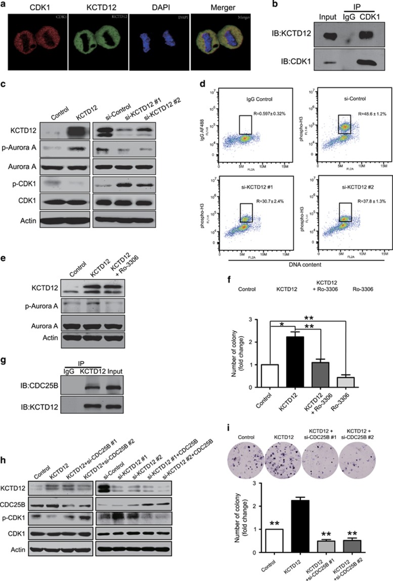
Figure 4.
KCTD12 interacts with CDC25B to promote G2/M transition and cell proliferation by dephosphorylating CDK1. (a) Confocal immunofluorescence analysis of KCTD12 and CDK1 localization in HeLa cells. KCTD12, green; CDK1, red; DAPI was used to label the nuclei; the cells presented are at metaphase. (b) HeLa cells were synchronized to M phase, and whole-cell lysates were immunoprecipitated with a CDK1 antibody, and then KCTD12 expression was detected by western blotting. (c) HeLa cells were transfected with KCTD12-expressing plasmid (1 μg, 24 h, left panel) or the si-KCTD12 (100 nM, 24 h, right panel), and p-Aurora A, Aurora A, p-CDK1 and CDK1 expression levels in the experimental group were compared with those in the control group by western blotting. Actin was included as a loading control. (d) Knockdown of KCTD12 decreased the percentage of M phase cells. The cells labeled with IgG AF488 were used as negative control. (e) Inhibiting CDK1 activity with Ro-3306 (30 nM, 24 h) attenuated the effect of KCTD12 (1 μg, 24 h) on Aurora A phosphorylation. (f) Ro-3306 decreased KCTD12-induced cell proliferation. HeLa cells were treated with Ro-3306 (30 nM, 24 h) or transfected with KCTD12-expressing plasmid (1 μg, 24 h) alone, or the combination, and colony-formation ability was compared. (g) The interaction between KCTD12 and CDC25B was examined by Co-IP assay in the HeLa cells synchronized at M phase. (h) KCTD12-expressing plasmid (1 μg) or vector control were co-transfected into HeLa cells with the siRNA against CDC25B (100 nM) or the si-Control (left panel); on the contrary, the siRNA against KCTD12 (100 nM) or si-Control was transfected into HeLa cells with or without CDC25B-expressing plasmid (right panel). Cells were harvested 24 h after transfection, and expression levels of KCTD12, CDC25B, p-CDK1 and CDK1 were determined by western blotting. (i) HeLa cells were co-transfected with KCTD12-expressing plasmid or vector control and si-CDC25B or si-Control, and the ability of the cells to form colonies was compared by colony formation assay. Bars, s.d.; *P<0.05; **P<0.01; ***P<0.001 compared with cells transfected with the KCTD12-expressing plasmid.

