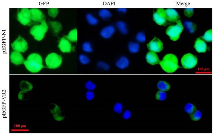
Figure 4.
Subcellular localization of LvVEGFR2 in mammalian 293T cells. The plasmid pEGFP-VR2, which was constructed based on the plasmid pEGFP-N1, contained an N-terminal IgK_secretion tag, the full length of enhanced green fluorescent protein (EGFP), and a C-terminal amino acid sequence including the predicted TM of LvVEGFR2 and its flanking sequence. The plasmid pEGFP-VR2 and control plasmid pEGFP-N1 were transfected into 293T cells and the fluorescence signals were detected, respectively. GFP showed fluorescence signals generated by expressed EGFP protein. DAPI showed signals generated by stained nucleus. Merge showed the merged signals of GFP and DAPI.

