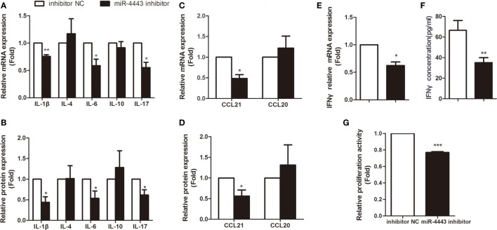
Figure 3.
Inhibiting miR-4443 expression in CD4+ T cells isolated from untreated Graves’ disease (uGD) patients downregulated T cell function and proliferation. CD4+ T cells isolated from GD patients were transfected with inhibitor negative controls or miR-4443inhibitors. Twelve hours after transfection, cells were stimulated with anti-CD3 and anti-CD28 antibodies. (A,C) Relative mRNA expression levels of interleukins (IL-1β, IL-4, IL-6, IL-10, IL-17) and chemokines (CCL21, CCL20) in CD4+ T cells. (B,D) Relative protein levels of interleukins (IL-1β, IL-4, IL-6, IL-10, IL-17) and chemokines (CCL21, CCL20) in the medium of cultured CD4+ T cells. (E,F) Relative mRNA expression and protein levels of IFN-γ. (G) Relative proliferation activity of cultured CD4+ T cells. Bars show the mean ± SD of three independent experiments. *P < 0.05; **P < 0.01; ***P < 0.001.

