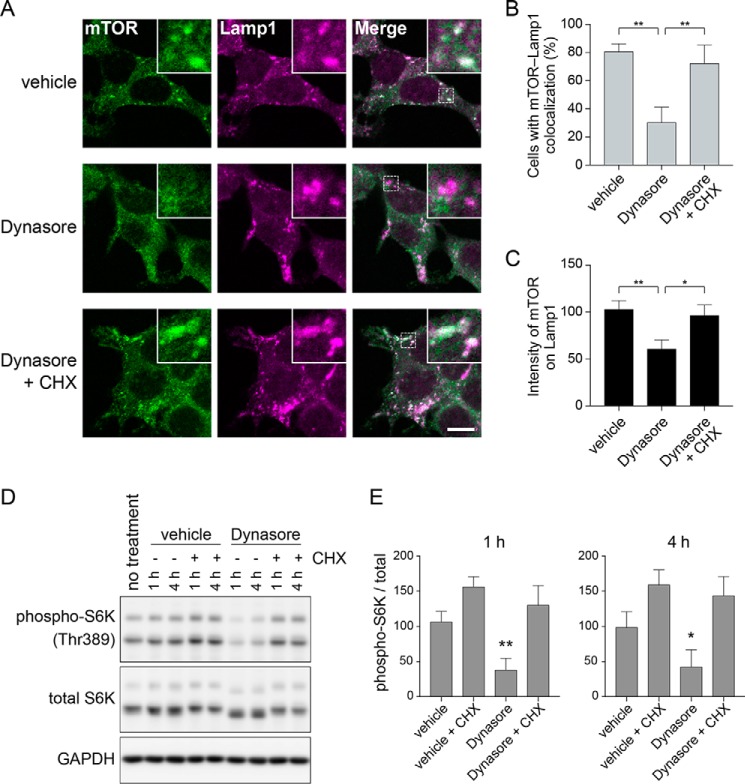
Figure 4.
Lysosomal localization and activation of mTORC1 are prevented by endocytosis inhibition and rescued by CHX. A, 293T cells were treated for 1 h with 0.2% DMSO, 80 μm Dynasore, or 80 μm Dynasore and 10 μg/ml of CHX, fixed, and stained with anti-mTOR and anti-Lamp1 antibodies. The results shown are representative of three independent experiments. Scale bar = 10 μm. B, quantification of cells that exhibit lysosomal localization of mTOR by analysis of image data in A. Each bar indicates mean ± S.D. of three independent experiments. **, p < 0.01. C, quantification of fluorescence intensity of mTOR on lysosomes by analysis of image data in A. Each bar indicates mean ± S.D. of three independent experiments. *, p < 0.05; **, p < 0.01. D, 293T cells were treated for 1 or 4 h with 0.2% DMSO, 10 μg/ml of CHX, 80 μm Dynasore, or 80 μm Dynasore and 10 μg/ml of CHX. E, quantification of immunoblot data in D. Each bar indicates mean ± S.D. of four independent experiments. *, p < 0.05; **, p < 0.01 compared with the vehicle treatment group.

