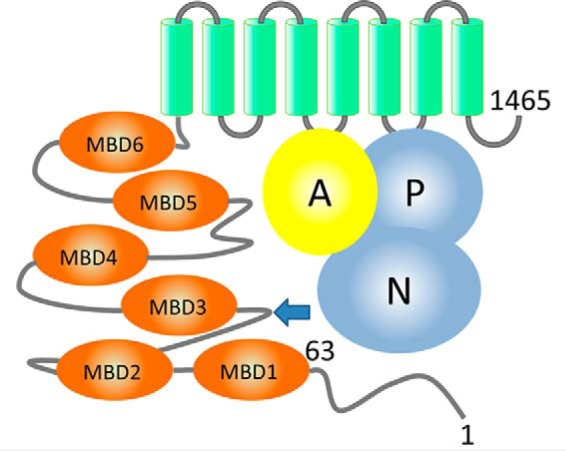
Figure 1.

Domain organization of ATP7B. ATP7B includes the N-terminal peptide (ATP7B1–63, residues labeled), six metal-binding domains (MBD1–MBD6, orange), and eight transmembrane helices (green). The N and P domains (blue) together hydrolyze ATP, with participation of the A domain (yellow). The arrow indicates the site of the spontaneous ATP7B proteolysis in the cell.
