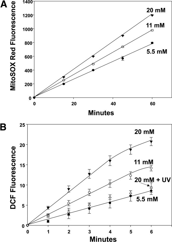
Figure 5.
Reactive species production by neutrophils exposed to 5.5, 11, or 20 mm glucose. A, murine neutrophils (5.5 × 10⁵/ml PBS containing 1 mm CaCl2, 1.5 mm MgCl2, and 5.5 mm glucose) were first incubated with 5 μm MitoSOX Red for 10 min, and then washed and resuspended in the buffer with different glucose concentrations as shown for up to 1 h. B, cells were incubated with the buffer shown for 2 h, and then 10 μm DCF-DA was added, and fluorescence was monitored. Note that when aliquots of cells were incubated in the different glucose concentrations for only 30 min or 1 h, DCF-DA was added, and the same fluorescence rates were measured as for incubations lasting 2 h. This method was chosen because, as discussed under “Experimental procedures,” DCF can autocatalyze its own oxidation (79). Values are mean ± S.E., n = 5 for each point. Using the same methods, effects of various inhibitors and siRNA depletion of cells are shown in Table 1.

