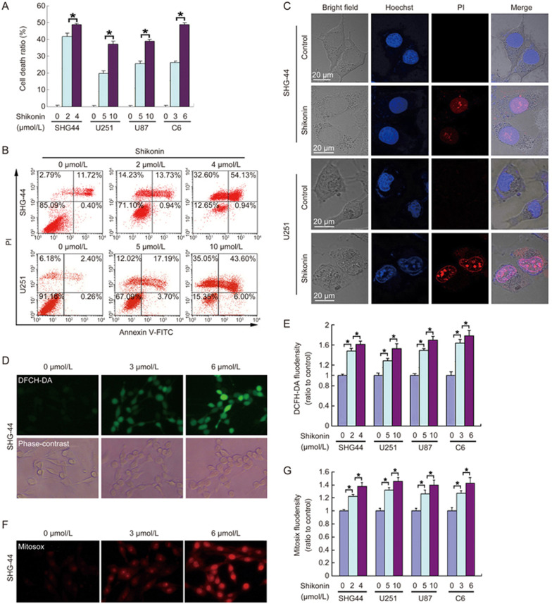
Figure 1.
Shikonin-induced necrosis in glioma cells was accompanied by an overproduction of intracellular ROS and mitochondrial superoxide. (A) The LDH release assay showed that shikonin induced glioma cell death in a concentration-dependent manner. (B) Flow cytometry with Annexin V/PI double staining proved that shikonin did not cause obvious increases in the percentage of Annexin V+/PI- (early apoptosis) cells but significantly increased the PI+ or Annexin V+/PI+ cells at 3 h of incubation. (C) Confocal microscopy with Hoechst 33342 and PI dual staining showed that shikonin did not induce chromatin condensation or nuclear fragmentation. (D) Representative phase-contrast and fluorescence microscopic images of SHG-44 glioma cells incubated with the ROS probe DCFH-DA at 2 h of incubation with shikonin (20×). (E) Statistical analysis of the fluorescence density detected by DCFH-DA demonstrates that shikonin induces an overproduction of intracellular ROS in a dosage-dependent manner. (F) Representative fluorescence microscopic images of the SHG-44 cells that were stained with the mitochondrial superoxide probe Mitosox Red at 2 h of incubation with shikonin (20×). (G) Statistical analysis of the red fluorescence density detected by Mitosox Red proves that the excessive generation of intracellular mitochondrial superoxide caused by shikonin was dependent on the shikonin concentration. The values are expressed as the mean±SD (n=5 per group). *P<0.01.

