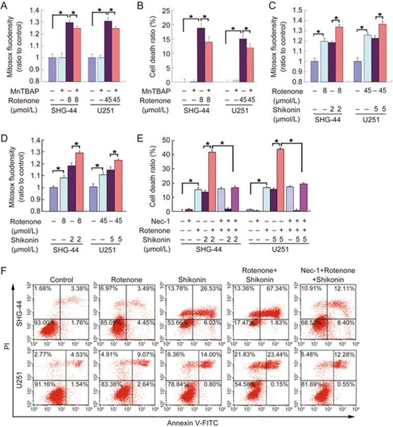
Figure 4.
ROS regulated shikonin-induced necrosis in glioma cells. (A) Statistical analysis of the fluorescence density detected by Mitosox Red proved that 4 h of incubation with rotenone resulted in an excessive generation of mitochondrial superoxide, which was significantly prevented by pretreatment with 40 μmol/L MnTBAP for 1 h. (B) The LDH release assay showed that MnTBAP inhibited rotenone-induced cell death in SHG-44 and U251 glioma cells. (C) Statistical analysis of the fluorescence density detected by Mitosox Red demonstrated that pretreatment with rotenone for 2 h enhanced the generation of mitochondrial superoxide caused by 2 h of incubation with shikonin. (D) Statistical analysis of the fluorescence density detected by DCFH-DA proved that pretreatment with rotenone for 2 h improved the increased intracellular ROS levels induced by 2 h of treatment with shikonin. (E) The LDH release assay showed that rotenone augmented shikonin-induced glioma cell death, which was prevented by Nec-1. However, Nec-1 did not prevent glioma cell death induced by rotenone alone. (F) Flow cytometry with Annexin V/PI double staining showed that rotenone enhanced shikonin-induced necrosis in glioma cells, but this enhancement effect was blocked by Nec-1. The values are expressed as the mean±SD (n=5 per group). *P<0.01 versus control group.

