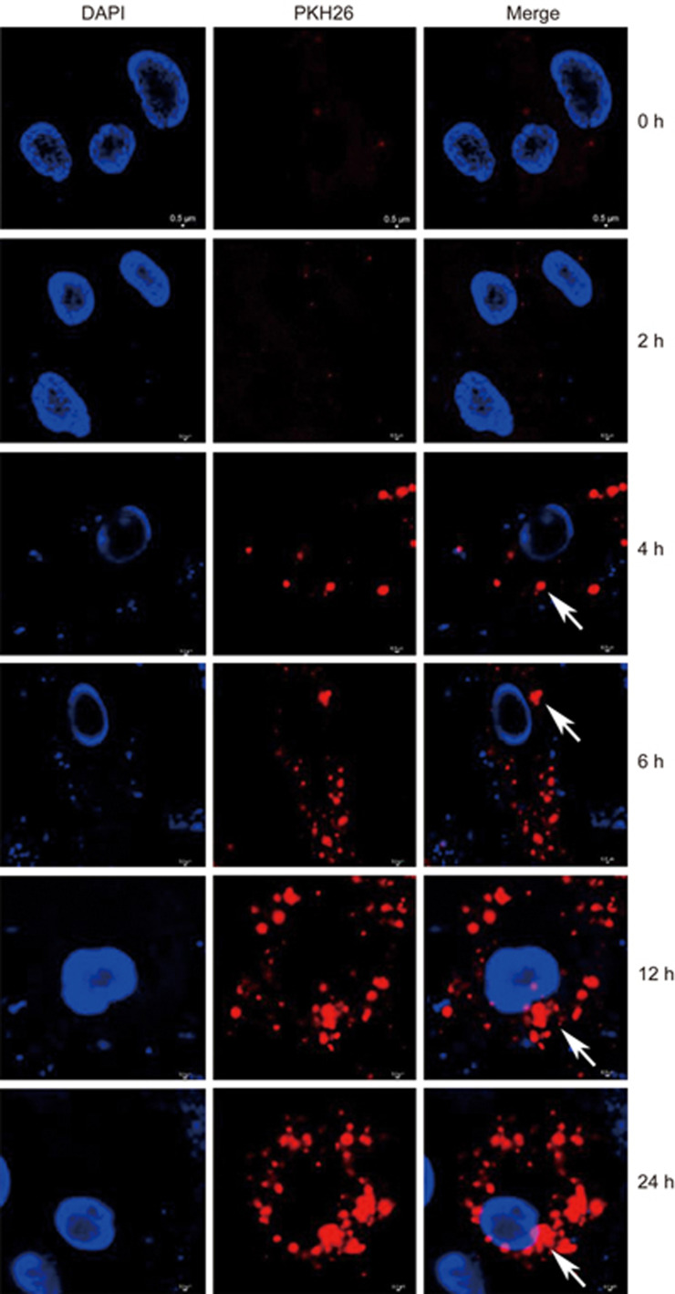
Figure 1.
Incorporation of K562-MVs into BM-MSCs. BM-MSCs were grown in 96-well plates for 24 h, and then PKH26-stained K562-MVs at 400 ng/mL were added. After 2, 4, 6, 12, 18 and 24 h of co-culture, the incorporation of K562-MVs into BM-MSCs with nuclei counterstained by DAPI was observed. Arrows show PKH26-stained K562-MVs (red) within the cytoplasm of BM-MSCs, around DAPI-labeled nuclei (blue), indicating the incorporation of K562-MVs into BM-MSCs. Stained cells and MVs were imaged using a confocal microscope (NikonA1Si, Nikon, Tokyo, Japan) after treatment with an anti-fluorescein quencher.

