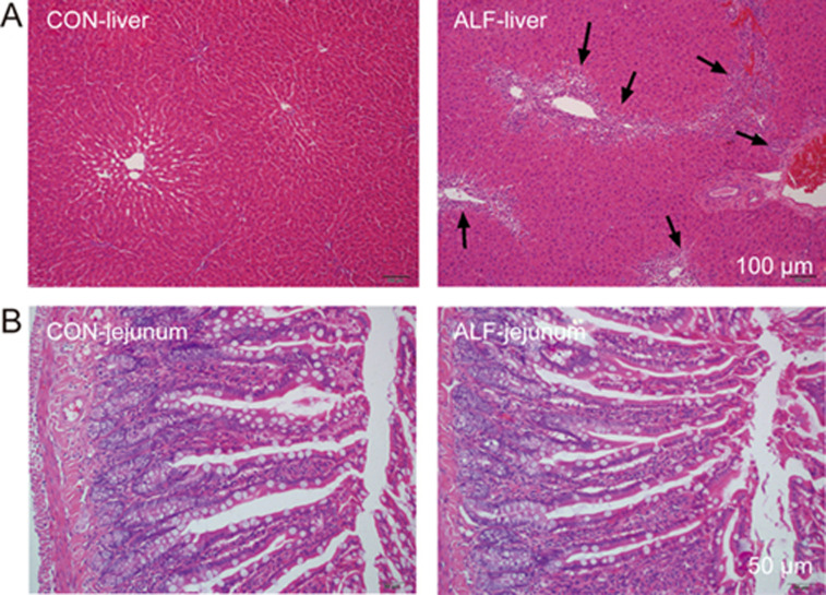
Figure 1.
Histological analyses of liver (A) and jejunum (B) from acute liver failure (ALF) rats and age-matched control (CON) rats. All sections were stained with hematoxylin-eosin (magnification, 200× for jejunum and 100× for liver). Scale bar represented 100 μm for liver and 50 μm for jejunum, respectively. Arrows indicate vacuolation and inflammatory cell infiltration.

