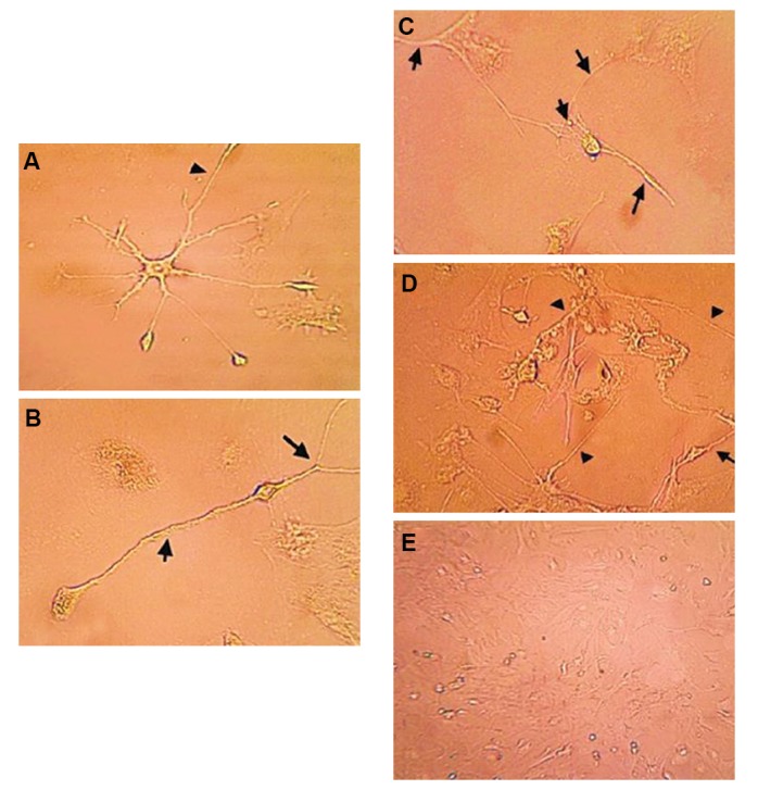
Fig.3.

Bone marrow mesenchymal stem cells (BM-MSCs) after 7 days in culture with the addition of 10% pre- and postnatal cerebrospinal fluid (CSF) photographed with phase-contrast optics. A. Culture with E19 CSF (×400), B. Culture with E20 CSF (×400), C. Culture with P1 CSF (×400), D. Basic fibroblast growth factor (b-FGF, positive control) (×400), and E. Control group (×200) without pre- and postnatal CSF. There was significantly greater neural differentiation of BM-MSCS cultured in CSFsupplemented medium from E19 compared to E20 and P1.
