
Fig.6.
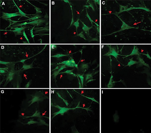
Cerebrospinal fluid (CSF) induced neuronal differentiation in the bone marrow mesenchymal stem cells (BM-MSCs). A. β-III tubulin expression in BMMSCs cultured with 10 ng/ml basic fibroblast growth factor (b-FGF) (×400), B. 10% E19 CSF (×400), C. 10% E20 CSF (×400), D. 10% P1 CSF (×400), E. MAP-2 expression in BM-MSCs cultured with 10 ng/ml b-FGF (×400), F. 10% E19 CSF (×400), G. 10% E20 CSF (×400), H. 10% P1 CSF (×400), and I. Control (×200) cultures without added CSF cells that show no stain when primary antibody was omitted. Red arrows indicate positive expression of β-III tubulin and MAP-2 in neuronlike cells and neurites. The highest expression level for these markers was detected in E19.