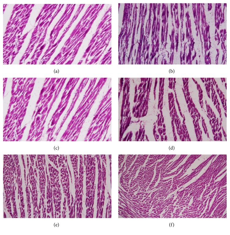
Figure 11.
Histopathological photomicrographs of heart tissue (6100x magnification; scale bar: 20 μm). (a) Normal control rats showed normal cardiac muscle fibers. (b) Diabetic control rats showed pathological changes including marked separation of cardiac muscle fibers and myocardial necrosis. (c–f) Glibenclamide- and GP-treated rats showed cardioprotection, with markedly increased areas of normal muscle fibers.

