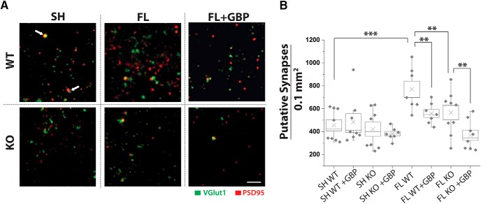
Figure 4.
GBP treatment or α2δ-1 deletion decreases FL-driven synaptogenesis. A, MIPs from of three optical sections of confocal images of VGlut1 (green) and PSD95 (red) collected at 100× from Layer V of WT and α2δ-1−/− (KO) FL and sham-injured animals, with vehicle or GBP treatment. White arrow indicates colocalization of VGLUT1 and PSD95, representing a site of synaptic contact. Scale bar = 500 nm. B, Box-whisker plot of number of synapses (colocalization of VGlut1/PSD95) per 1.00 mm3 per MIP, **α = 0.01 and ***α = 0.001 (Holm-Bonferroni multiple-comparison correction).

