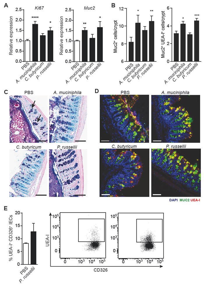
Figure 3. P. russellii promotes goblet cell differentiation and function in vivo.

Mice were orally gavaged with PBS, A. muciniphila, C. butyricum, or P. russellii every other day for 2 weeks and the distal large intestine was harvested for analysis.
(A) Quantitative RT-PCR showing expression of proliferation marker Ki67 (****P < 0.0001; *P = 0.0105) and goblet cell-derived mucin 2 (Muc2; **P = 0.0020; *P = 0.0275), relative to Gapdh. n = 10 per group. Data pooled from 2 independent experiments.
(B) Quantification of Muc2+ (**P = 0.0088; *P = 0.0418) and Muc2+UEA-I+ (***P = 0.0009; *P = 0.0184) goblet cell number per crypt in the distal colon, n = 5 per group.
(C) Representative alcian blue and periodic acid-Schiff (AB/PAS)-stained distal colon sections showing goblet cell staining within the mucosa. Goblet cells are indicated by black arrows. Scale bar, 100 μm.
(D) Representative epifluorescence staining for goblet cells in the distal colon using an antibody against Muc2 (green) and the lectin UEA-I (red) with DAPI (blue) as the counter stain. Scale bar, 100 μm.
(E) Intracellular flow cytometry analysis of isolated colonic epithelial cells (CD326+) shows increased presence of UEA-1+CD326+ double-positive cells in P. russellii-treated animals. The lectin UEA-1 binds α-1,2 fucose. Significance determined using Student’s t-test and expressed as mean + SEM.
See also Figure S2.
