
Figure 2.
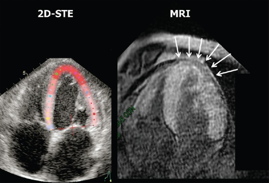
Comparison between the two-dimensional speckle-tracking echocardiography functional image of the left ventricle and contrast magnetic resonance imaging. The magnetic resonance imaging T1 late gadolinium enhancement (fast-gradient-echo inversion-recovery) image was acquired 10 min after bolus intravenous injection of 0.15 mmol/kg body weight of gadolinium-diethylenetriamine pentaacetic acid (gadobutrol, Schering, Germany), followed by saline flush. The inversion time was adapted to null normal myocardium. The arrows on the magnetic resonance imaging image indicate an apical area with relative absence of contrast uptake, which corresponds to the red color at the apex of the left ventricle on the two-dimensional speckle-tracking echocardiography functional image