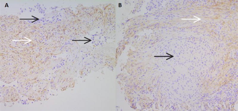
Figure 3.
A. Results of PDGFRα IHC in a core biopsy of an adenocarcinoma. The tumor is negative, and moderate cell membrane and cytoplasmic staining of stromal cells can be seen.
B. Results of PDGFRα IHC in a core biopsy of a squamous cell carcinoma. The tumor is negative, and surrounding stromal fibroblasts show light to moderate cell membrane and cytoplasmic staining, beginning to intersperse among lymphocytes in the lower right corner. Both images are at 100× magnification.
In both A and B, black arrows identify tumor, while white arrows identify positively staining tumor-associated stroma.

