. 2017 Aug 29;8(11):7368–7373. doi: 10.1039/c7sc02420g

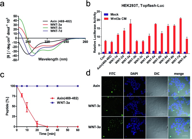
Fig. 3. (a) CD spectra of the stapled peptides. The peptides were dissolved in PBS buffer at a final concentration of 50 μM. The percent helicity was calculated based on the [θ]222 value. (b) HEK293T cells transfected with Topflash-luciferase were treated with Wnt3a conditioned medium (CM) and target peptides (40 μM) at the indicated concentrations for 12 h and then harvested for luciferase measurement. Con = non-specific control. (c) Proteolytic stability of the peptides (Axin (469–482) vs. WNT-3a) in the α-chymotrypsin solution (5 ng μL–1 in 50 mM PBS buffer, pH = 7.4) at a final concentration of 0.1 mM. Data points are displayed as the mean value SEM of duplicate independent experiments. The percent of residual peptide was monitored by analytical HPLC. (d) Confocal microscopic images of FITC–β-Ala–Axin, -3a, and -3c treated HEK 293T/17 cells. The cells were incubated with FITC–β-Ala–peptide (40 μM) for 16 h and then washed with PBS twice before fixation for confocal microscopy. Scale bar: 50 μm.

Fig. 3