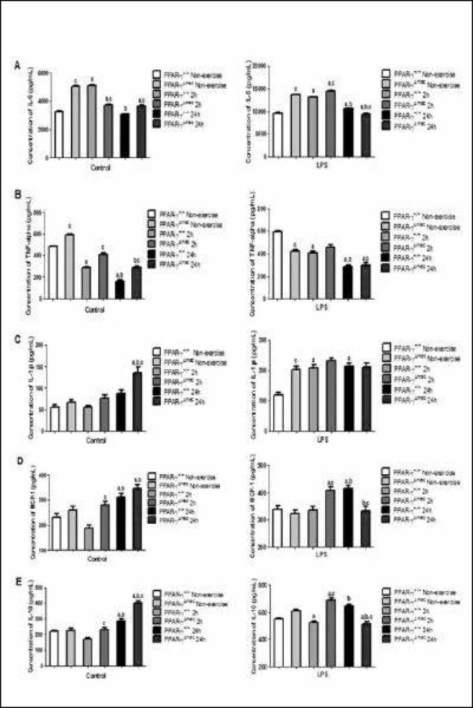
Fig. 1.
Secretion of (A) interleukin 6 (IL-6), (B) tumor necrosis factor-α (TNFα), (C) interleukin- one beta (IL-1beta), (D) monocyte chemoattractant protein-1 (MCP-1) and (E) interlukin-10 (IL-10) in cultured peritoneal macrophages from PPAR-γ(+/+) and PPAR-γΔmac mice. Cells from the groups: non-exercise, 2h and 24h after one bout of moderate exercise protocol. Cells were cultured in 24-well dishes (5 × 10⁵ cells/well) and stimulated with LPS (2,5 ng/ml) or not for 24h. The concentration of citokynes in the culture media was determined by ELISA. Data are presented as mean ± SEM. a = different from control; b = different from 2h; c = different between genotype (One-way ANOVA followed by Bonferroni; p < 0.05).

