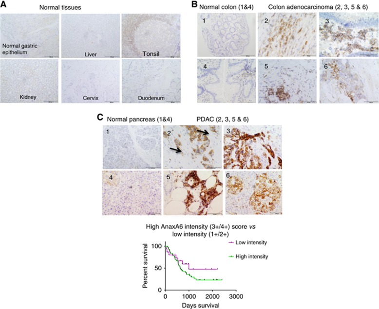
Figure 4.
IHC analysis with MAb 9E1 demonstrates that AnxA6 shows limited expression in normal and highly proliferating tissues (A) with strong membrane AnxA6 expression observed in colon adenocarcinoma (B) and PDAC (C). High AnxA6 expression was significantly associated with the presence of PNI (P=0.0001) and with tumours exhibiting tumour budding (P=0.0082) in PDAC and showed a weak correlation with outcome (P=0.2242) in this 57 PDAC cohort.

