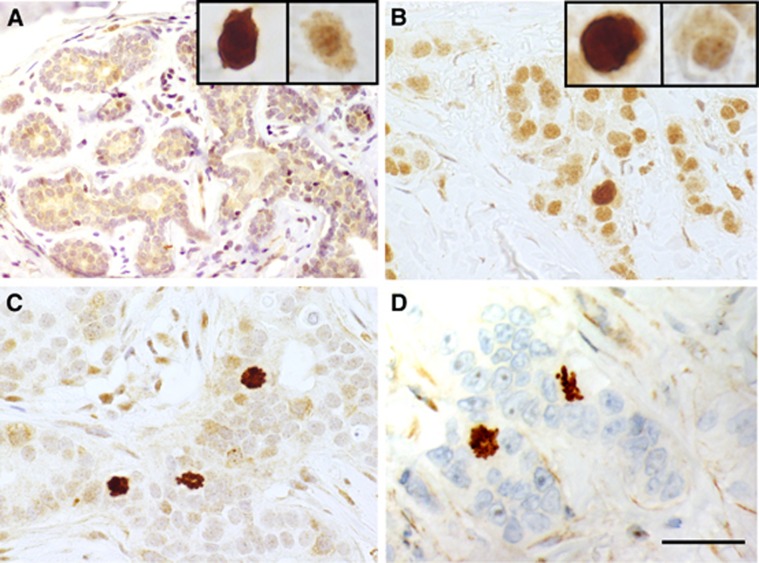
Figure 1.
Examples of separase immunoexpression in invasive breast carcinomas and benign breast epithelium. Separase positivity was observed as two distinct staining patterns in the cancer cells, diffuse separase expression in the cytoplasm and nucleus, and mitotic separase. Benign breast tissue showed a clear diffuse separase expression and only single separase-positive mitoses reflecting the low proliferation rate in normal breast epithelium (A). In breast carcinomas representing luminal (B), HER2-amplified (C) and triple-negative breast carcinomas (D), the two staining patterns occurred inversely related so that strong diffuse separase-positivity was associated with low mitotic separase-expression and vice versa. (magnification × 400, space bar 100 μm). A full colour version of this figure is available at the British Journal of Cancer journal online.

