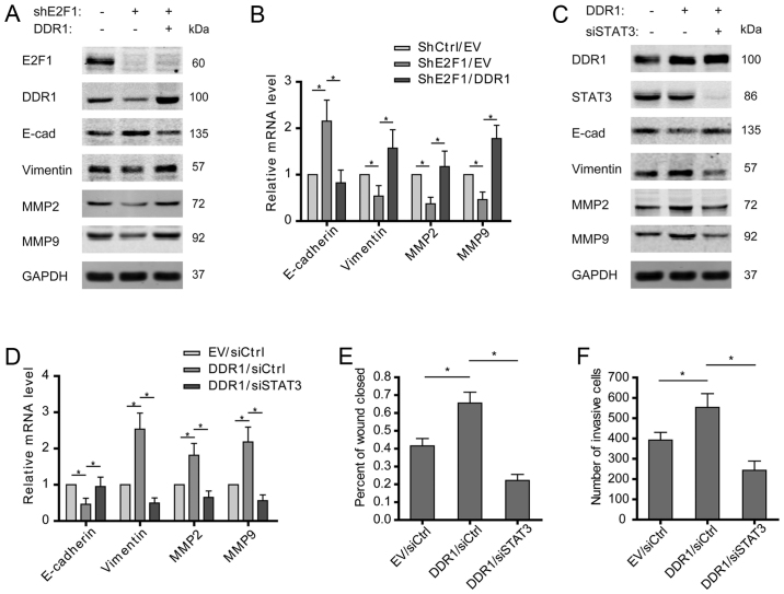
Figure 5.
E2F1/DDR1/STAT3 axis drives the EMT of osteosarcoma cells. (A and B) Shscramble or shE2F1-U2OS cells were transfected with empty vector and DDR1 plasmids, and the expression of E-cadherin, vimentin, MMP2, and MMP9 was examined. (C and D) U2OS cells were transfected with empty vector/DDR1 plasmids and siscramble/siSTAT3. The expression of E-cadherin, vimentin, MMP2, and MMP9 was examined. (E and F) Cell migration or invasion activity was examined by wound healing or Transwell assay. Wound areas or invasive cells were quantified. (B and D-F) Data shown are mean ± SEM, each performed in triplicate. *P<0.05.

