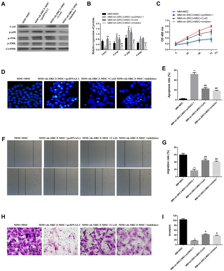
Figure 5.
MAPK pathway is involved in promoting the proliferation and migration of RPMI-8226 cells regulated by Cx43. The RPMI-8226 cells were transfected with either pcDNA3.1 or pcDNA3.1-Cx43 for 48 h. These cells were treated with 5 µM MAPK inhibitor SB202190 for 24 h, and then co-cultured with either BMSCs or sh-SRC3-MSC. (A) Western blots analyzed the protein level of Cx43, phosphorylated ERK (pERK), p38 (p-p38) and JNK (p-JNK) in RPMI-8226 cells. (B) Densitometry plot of results from (A). The relative expression levels were normalized to GAPDH. (C) Cell proliferation analysis of RPMI-8226 cells after being co-cultured for 48 h using the CCK-8 assay. (D) Hoechst foci staining for co-cultured RPMI-8226 cells. (E) The cells that stained positive for Hoechst staining were counted. (F and G) Scratch-wound healing assay was used to assess the migration potential of RPMI-8226 cells after being co-cultured for 48 h. The wound closure rate was calculated at 24 h using a phase contrast microscope. (H) Transwell migration assay assessed the change of migration potential of RPMI-8226 cells after being co-cultured for 48 h. Representative images of migrated cells are shown. (I) Relative numbers of migrated cells in the Transwell assay under a phase contrast microscope. Data represent three independent experiments (average and SEM of triplicate samples). *P<0.05, **P<0.01 vs. control; #P<0.05, ##P<0.01 vs. MM+sh-SRC3-MSC+pcDNA3.1.

