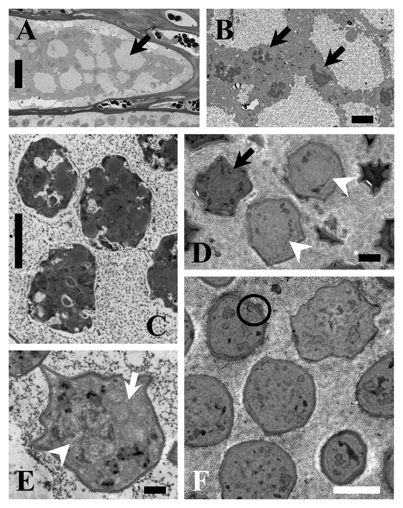
Figure 3.
TEM images of M. braseltonii. A: Cell filled with a lobose plasmodium (Arrow). Scale bar: 20 μm. B: Details of the multiple, dividing nuclei (arrows) distinctive for growing plasmodia. Scale bar: 2 μm. C: Plasmodium cleaving into resting spores. The individual cells are already organised but the cell wall is not yet visible. Scale bar: 2 μm. D: Maturing resting spores, which are still slightly irregular in shape (arrowheads), but the multi-layered cell wall is already visible in some of them (arrow). Scale bar: 1 μm. E: Detail showing the nucleus (arrowhead) and mitochondria (arrow) of a developing resting spore. 500 nm. F: Ripe resting spores with the characteristic multi-layered cell wall (circle). Scale bar: 2 μm.

